An up-to-date list is available on Google Scholar.
2023
-

×
![]()
Zero-Shot Self-Supervised Joint Temporal Image and Sensitivity Map Reconstruction via Linear Latent Space
In Medical Imaging with Deep Learning – MIDL 2023
Fast spin-echo (FSE) pulse sequences for Magnetic Resonance Imaging (MRI) offer important imaging contrast in clinically feasible scan times. T2-shuffling is widely used to resolve temporal signal dynamics in FSE acquisitions by exploiting temporal correlations via linear latent space and a predefined regularizer. However, predefined regularizers fail to exploit the incoherence especially for 2D acquisitions.Recent self-supervised learning methods achieve high-fidelity reconstructions by learning a regularizer from undersampled data without a standard supervised training data set. In this work, we propose a novel approach that utilizes a self supervised learning framework to learn a regularizer constrained on a linear latent space which improves time-resolved FSE images reconstruction quality. Additionally, in regimes without groundtruth sensitivity maps, we propose joint estimation of coil-sensitivity maps using an iterative reconstruction technique. Our technique functions is in a zero-shot fashion, as it only utilizes data from a single scan of highly undersampled time series images. We perform experiments on simulated and retrospective in-vivo data to evaluate the performance of the proposed zero-shot learning method for temporal FSE reconstruction. The results demonstrate the success of our proposed method where NMSE and SSIM are significantly increased and the artifacts are reduced.
-
Latent Signal Models: Learning Compact Representations of Signal Evolution for Improved Time-Resolved, Multi-Contrast MRI
Magnetic Resonance in Medicine, 2023
Purpose: Training auto-encoders on simulated signal evolution and inserting the decoder into the forward model improves reconstructions through more compact, Bloch-equation-based representations of signal in comparison to linear subspaces. Methods: Building on model-based nonlinear and linear subspace techniques that enable reconstruction of signal dynamics, we train auto-encoders on dictionaries of simulated signal evolution to learn more compact, non-linear, latent representations. The proposed Latent Signal Model framework inserts the decoder portion of the auto-encoder into the forward model and directly reconstructs the latent representation. Latent Signal Models essentially serve as a proxy for fast and feasible differentiation through the Bloch-equations used to simulate signal. This work performs experiments in the context of T2-shuffling, gradient echo EPTI, and MPRAGE-shuffling. We compare how efficiently auto-encoders represent signal evolution in comparison to linear subspaces. Simulation and in-vivo experiments then evaluate if reducing degrees of freedom by inserting the decoder into the forward model improves reconstructions in comparison to subspace constraints. Results: An auto-encoder with one real latent variable represents FSE, EPTI, and MPRAGE signal evolution as well as linear subspaces characterized by four basis vectors. In simulated/in-vivo T2-shuffling and in-vivo EPTI experiments, the proposed framework achieves consistent quantitative NRMSE and qualitative improvement over linear approaches. From qualitative evaluation, the proposed approach yields images with reduced blurring and noise amplification in MPRAGE shuffling experiments. Conclusion: Directly solving for non-linear latent representations of signal evolution improves time-resolved MRI reconstructions through reduced degrees of freedom.
-

×
![]()
Stochastic-offset-enhanced restricted slice excitation and 180° refocusing designs with spatially non-linear ∆B0 shim array fields.
Magnetic Resonance in Medicine, 2023
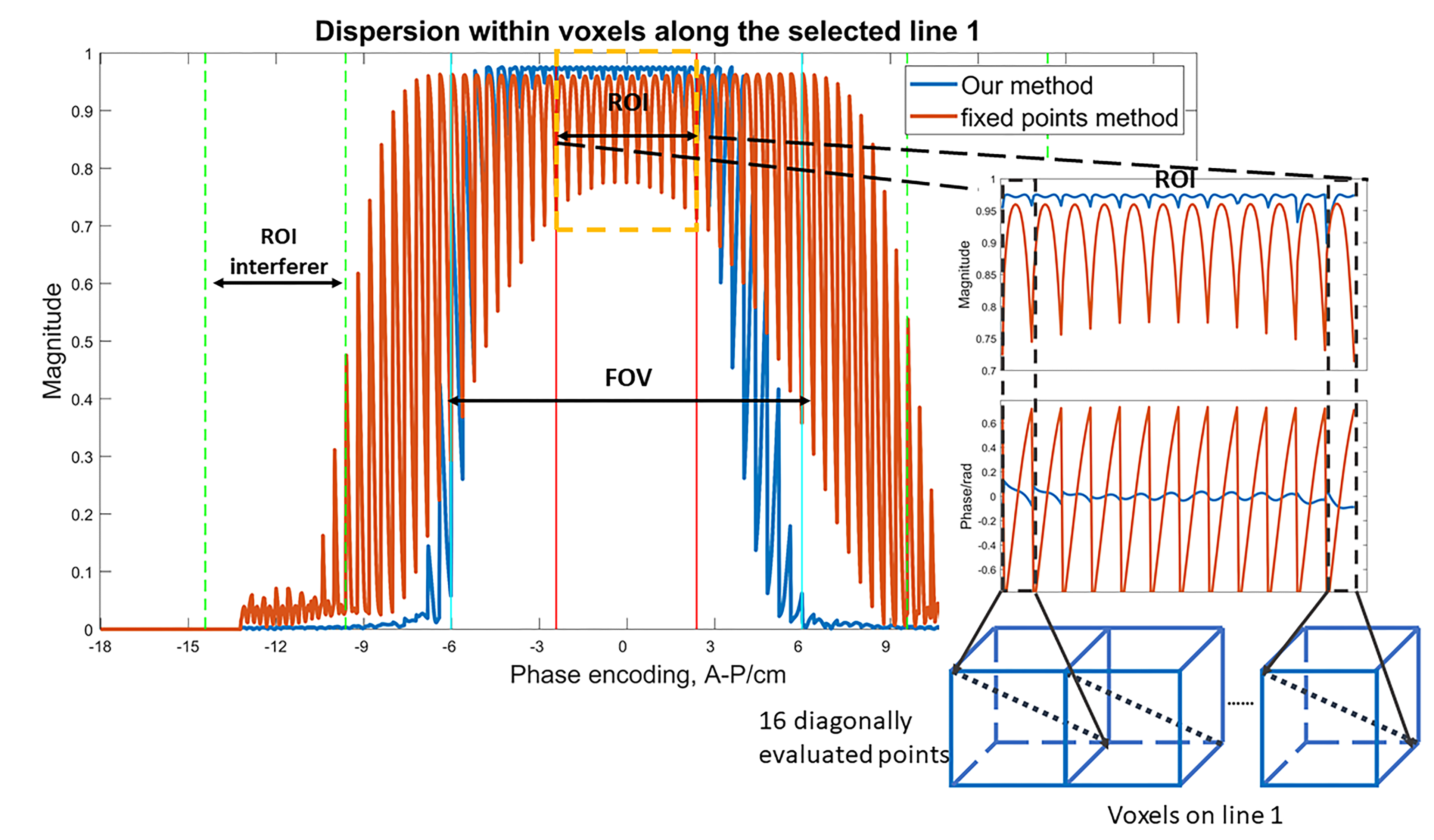
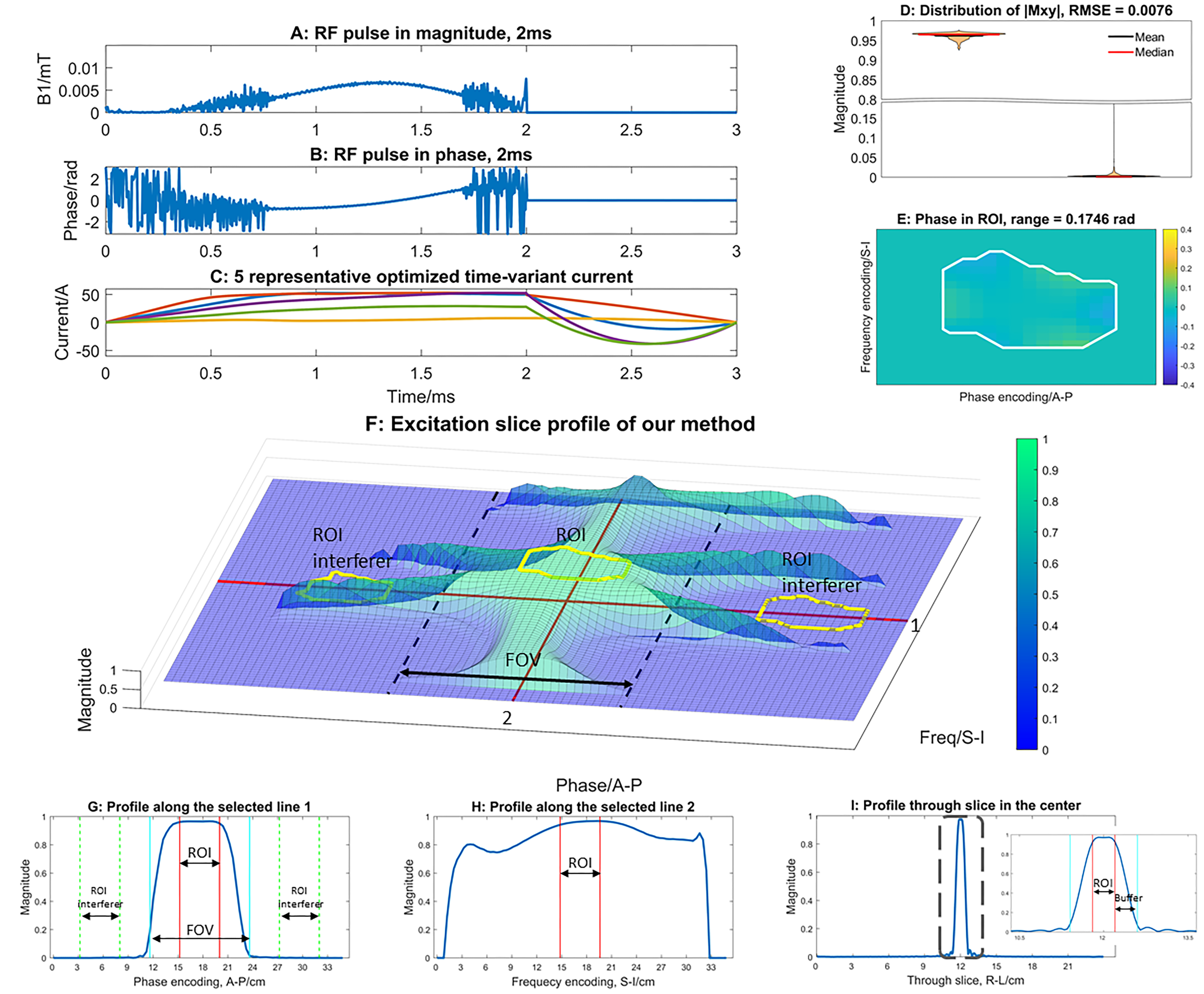
AbstractPurpose:Developing a general framework with a novel stochastic offset strat-egy for the design of optimized RF pulses and time-varying spatially non-linearΔB0shim array fields for restricted slice excitation and refocusing with refinedmagnetization profiles within the intervals of the fixed voxels.Methods:Our framework uses the decomposition property of the Blochequations to enable joint design of RF-pulses and shim array fields for restrictedslice excitation and refocusing with auto-differentiation optimization. Blochsimulations are performed independently on orthogonal basis vectors, Mx, My,and Mz, which enables designs for arbitrary initial magnetizations. Require-ments for refocusing pulse designs are derived from the extended phase graphformalism obviating time-consuming sub-voxel isochromatic simulations tomodel the effects of crusher gradients. To refine resultant slice-profiles becauseofvoxelwiseoptimizationfunctions,weproposeanalgorithmthatstochasticallyoffsets spatial points at which loss is computed during optimization.Results:We first applied our proposed design framework to standardslice-selective excitation and refocusing pulses in the absence of non-linearΔB0shim array fields and compared them against pulses designed with Shinnar-LeRoux algorithm. Next, we demonstrated our technique in a simulated setup offetal brain imaging in pregnancy for restricted-slice excitation and refocusing ofthe fetal brain.Conclusions:Our proposed framework for optimizing RF pulse andtime-varying spatially non-linearΔB0shim array fields achieve high fidelityrestricted-slice excitation and refocusing for fetal MRI, which could enablezoomed fast-spin-echo-MRI and other applications.
2022
-

×
![]()
Selective RF excitation designs enabled by time-varying spatially non-linear ∆B0 fields with applications in fetal MRI
Molin Zhang, Nicolas Arango, Jason P Stockmann, Jacob White, and
1 more author
Magnetic Resonance in Medicine, 2022
Purpose: To demonstrate, through numerical simulations, novel designs of spatially selective radiofrequency (RF) excitations of the fetal brain by both a restricted 2D slice and 3D inner-volume selection. These designs exploit a single channel RF pulse, conventional gradient fields, and the spatially non-linear ΔB0 fields of a multi-coil shim array, using an auto-differentiation optimization algorithm. Methods: The design algorithm jointly optimizes the RF pulse and the time varying ΔB0 fields, which is produced by a 64-channel multi-coil ΔB0 body array to augment the RF and the linear gradient fields, using an auto-differentiation approach. Two design targets were specified, one a 4-mm thick slice with a limited in-slice extent in one dimension (“restricted slice”), and the other a 3D inner-volume selection encompassing the fetal brain (“inner volume”). The RF duration was limited to 2 ms for the restricted slice excitation and 6 ms for the inner-volume excitation. Results: Excitation profiles were achieved for both the restricted slice excitation task (one-minus-minimum magnitude, 8%) within the region of interest (ROI) and (maximum-minus-zero magnitude, 8%) in the suppressed regions and the fetal brain volume excitation task (13% and 9%, respectively). Conclusions: The proposed joint design of RF and time-varying, spatially nonlinear ΔB0 fields achieves the target excitation profiles with short RF pulse durations and demonstrates the potential to enhance fetal MRI with multi-channel body shim arrays.
-
Zero-Shot Self-Supervised Learning for 2D T2-shuffling MRI Reconstruction
In International Society for Magnetic Resonance in Medicine 30th Scientific Meeting, 2022
-
Selective Refocusing Pulse Design via Time-varying Nonlinear Shim Array Fields and RF Pulse with Decomposition Property
In International Society for Magnetic Resonance in Medicine 30th Scientific Meeting, 2022
-
An Automated Pose and Motion Estimation Pipeline in Dynamic 3D Fetal MRI
In International Society for Magnetic Resonance in Medicine 30th Scientific Meeting, 2022
2021
-
Inner Volume Excitation via Joint Design of Time-varying Nonlinear Shim-array Fields and RF Pulse
In International Society for Magnetic Resonance in Medicine 29th Scientific Meeting, 2021
2020
-

×
![]()
Enhanced Detection of Fetal Pose in 3D MRI by Deep Reinforcement Learning with Physical Structure Priors on Anatomy
In Medical Image Computing and Computer Assisted Intervention – MICCAI 2020
Fetal MRI is heavily constrained by unpredictable and substantial fetal motion that causes image artifacts and limits the set of viable diagnostic image contrasts. Current mitigation of motion artifacts is predominantly performed by fast, single-shot MRI and retrospective motion correction. Estimation of fetal pose in real time during MRI stands to benefit prospective methods to detect and mitigate fetal motion artifacts where inferred fetal motion is combined with online slice prescription with low-latency decision making. Current developments of deep reinforcement learning (DRL), offer a novel approach for fetal landmarks detection. In this task 15 agents are deployed to detect 15 landmarks simultaneously by DRL. The optimization is challenging, and here we propose an improved DRL that incorporates priors on physical structure of the fetal body. First, we use graph communication layers to improve the communication among agents based on a graph where each node represents a fetal-body landmark. Further, additional reward based on the distance between agents and physical structures such as the fetal limbs is used to fully exploit physical structure. Evaluation of this method on a repository of 3-mm resolution in vivo data demonstrates a mean accuracy of landmark estimation 10 mm of ground truth as 87.3%, and a mean error of 6.9 mm. The proposed DRL for fetal pose landmark search demonstrates a potential clinical utility for online detection of fetal motion that guides real-time mitigation of motion artifacts as well as health diagnosis during MRI of the pregnant mother.
-
3D Fetal Pose Estimation with Adaptive Variance and Conditional Generative Adversarial Network
In Medical Ultrasound, and Preterm, Perinatal and Paediatric Image Analysis, 2020
Fetal motion is the dominant challenge to reliable performance and diagnostic quality of fetal magnetic resonance imaging (MRI). The fetus can move unpredictably and rapidly, leading to severe image artifacts. Consequently, MR acquisitions are largely limited to so-called single-shot techniques in an attempt to “freeze” fetal motion through fast imaging, while the problem due to motion occur between slices still exists. In this work, we propose a deep learning method for fetal pose estimation from MR volumes using the paradigm of conditional generative adversarial network which consists of two networks, a generator and a discriminator. The generator is responsible for estimating keypoint heatmaps from input MRI and the discriminator tries to learn the features of plausible fetal pose and distinguish ground-truth heatmaps from generated ones. With this adversarial training scheme, the generator can robustly produce realistic heatmaps for fetal pose inference. Besides, we use adaptive variance to model the difference in intensity of motion of different keypoints. Evaluation shows that the proposed method can improve the performance of pose estimation in 3D MRI, achieving quantitatively an average error of 2.64 mm and 98.31% accuracy (with error less than 10 mm). The proposed method can process volumes with latency less than 300 ms, potentially enabling low-latency online tracking of fetal pose during MR scans.
-
Landmark detection of fetal pose in volumetric MRI via deep reinforcement learning
In International Society for Magnetic Resonance in Medecine 28th Scientific Meeting, 2020
2019
-

×
![]()
Fetal Pose Estimation in Volumetric MRI using a 3D Convolution Neural Network
In Medical Image Computing and Computer Assisted Intervention – MICCAI 2019
The performance and diagnostic utility of magnetic resonance imaging (MRI) in pregnancy is fundamentally constrained by fetal motion. Motion of the fetus, which is unpredictable and rapid on the scale of conventional imaging times, limits the set of viable acquisition techniques to single-shot imaging with severe compromises in signal-to-noise ratio and diagnostic contrast, and frequently results in unacceptable image quality. Surprisingly little is known about the characteristics of fetal motion during MRI and here we propose and demonstrate methods that exploit a growing repository of MRI observations of the gravid abdomen that are acquired at low spatial resolution but relatively high temporal resolution and over long durations (10–30 min). We estimate fetal pose per frame in MRI volumes of the pregnant abdomen via deep learning algorithms that detect key fetal landmarks. Evaluation of the proposed method shows that our framework achieves quantitatively an average error of 4.47 mm and 96.4% accuracy (with error less than 10 mm). Fetal pose estimation in MRI time series yields novel means of quantifying fetal movements in health and disease, and enables the learning of kinematic models that may enhance prospective mitigation of fetal motion artifacts during MRI acquisition.
-
Fetal pose estimation via deep neural network by detection of fetal joints, eyes, and bladder
In International Society for Magnetic Resonance in Medecine 27th Scientific Meeting, 2019


